 Digital image processing started to be used for marine
particles as early as the 1970 s as computer technology became powerful
enough and available to scientists. An approach to automatically recognize
diatoms for pollution monitoring was developed first in the early 1970 s.
(Cairns et al., 1972), and was refined to use spatially matched optical
filters, video and computer technology to determine particle shape
(Almeida and Eu, 1976), and was later simplified to use rotating spatial
filters (i.e. holograms) (Fujii et al., 1980). Automated recognition of
phytoplankton cell types by digital image analysis was also demonstrated
in the 1970 s (Uhlmann et al., 1978). An automated system for pattern
recognition of cells of the toxic algal Prorocentrum in Japan has been
described (Tsuji and Nishikawa, 1984). Similarly, an image recognition
system for zooplankton was described by Jeffries et al. that achieved
classification to major taxonomic groups (Jeffries et al., 1984).
Zooplankton (Rolke and Lenz, 1984) and detrital (Lenz, 1972) size spectra
have been measured by digital image analysis with optical microscopy.
Digital image processing started to be used for marine
particles as early as the 1970 s as computer technology became powerful
enough and available to scientists. An approach to automatically recognize
diatoms for pollution monitoring was developed first in the early 1970 s.
(Cairns et al., 1972), and was refined to use spatially matched optical
filters, video and computer technology to determine particle shape
(Almeida and Eu, 1976), and was later simplified to use rotating spatial
filters (i.e. holograms) (Fujii et al., 1980). Automated recognition of
phytoplankton cell types by digital image analysis was also demonstrated
in the 1970 s (Uhlmann et al., 1978). An automated system for pattern
recognition of cells of the toxic algal Prorocentrum in Japan has been
described (Tsuji and Nishikawa, 1984). Similarly, an image recognition
system for zooplankton was described by Jeffries et al. that achieved
classification to major taxonomic groups (Jeffries et al., 1984).
Zooplankton (Rolke and Lenz, 1984) and detrital (Lenz, 1972) size spectra
have been measured by digital image analysis with optical microscopy.
In situ cameras systems have become common and
widespread on remotely operated vehicles (ROVs). In the past decade the
Video Plankton Recorder (VPR) has been developed for the automated mapping
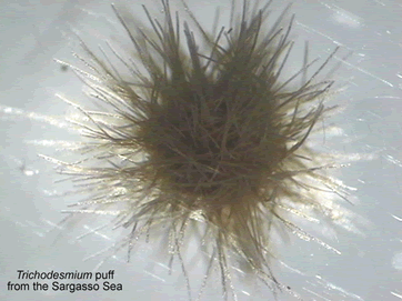
and analysis of zooplankton (Davis et al., 1992) and larger phytoplankton
colonies such as the Chaetoceros, Phaeocystis, and Rhizosolenia mats. In-situ
holographic cameras are being developed and tested to image volumes of
water to show spatial patterns at small scales (Craig et al. 2000; Katz et
al. 1999). Jaffe and Franks (1996) are developing an in-situ fluorescence
imaging system for studying small scale patterns of phytoplankton
distributions.
 Automated techniques for optical fluorescence
microscopy have also been developed for marine bacteria (Sieracki et al.,
1985) and protists (Sieracki and Viles, 1990). This work includes the
evaluation of threshold methods for segmenting images of fluorescing cells
(Sieracki et al., 1989a), (Viles and Sieracki, 1992), an algorithm for
calculating cell biovolume from 2-D images (Sieracki et al., 1989b), and
the accurate counting and sizing of cells from images (Sieracki and Viles,
1998). The largest remaining challenge in the image analysis of
fluorescence microscopy images is the recognition and classification of
particle types, especially in the nanoplankton samples that can contain
large amounts of detrital particles and can be confused with cells.
Automated techniques for optical fluorescence
microscopy have also been developed for marine bacteria (Sieracki et al.,
1985) and protists (Sieracki and Viles, 1990). This work includes the
evaluation of threshold methods for segmenting images of fluorescing cells
(Sieracki et al., 1989a), (Viles and Sieracki, 1992), an algorithm for
calculating cell biovolume from 2-D images (Sieracki et al., 1989b), and
the accurate counting and sizing of cells from images (Sieracki and Viles,
1998). The largest remaining challenge in the image analysis of
fluorescence microscopy images is the recognition and classification of
particle types, especially in the nanoplankton samples that can contain
large amounts of detrital particles and can be confused with cells.
More recently, work on automated pattern recognition of phytoplankton has
been done by Europeans (Culverhouse et al., 1996). This study compared
classification methods and human experts with a set of images of 23
dinoflagellates species from 4 genera. The best algorithm was a Radial
Basis Function neural network classifier that performed as well as the
experts (84% accurate). The self-learning neural network methods
outperformed the classical multivariate statistical approaches. This
classification system has also been used for loricate marine ciliates (Culverhouse
et al., 1994). The software, termed DiCANN (Dinoflagellate Categorisation
by Artificial Neural Network (Culverhouse et al., 2002)) is under
development for
commercialization, but is not yet available. If it becomes available
during this project and it is
affordable we will purchase it and test it with our images.
These results show that neural network algorithms can approach human
experts in categorizing dinoflagellates from field samples, however there
is much more work to be done. The test set was rather limited and the
images were digitized from film images, causing artifacts (Culverhouse et
al., 1996). One limitation of neural network classification solutions is
that, although they are powerful and often perform well, the nature of the
complex network created prevents a full understanding of how the input
features are weighted and used. Also the features chosen to be extracted
from the images may not be the best ones, and they were not systematically
tested. So there remains much work to be done.