In-situ marine particle imaging
Our priority application for the marine snow instrument is quantification
of vertically migrating algal mats in the oligotrophic North Pacific that
transport nitrate from below the thermocline to the surface. This
transport flux substantially impacts the cycling of nitrogen and overall
primary production rates. Images archived from recent field work wil be
used. A proposed application (proposal pending) involves determining the
size and abundance of large organic particles within the pervasive benthic
nepheloid layer that represent a rich, near-bottom food source and sites
of biogenic transformations of organic matter. Detrital particles and
zooplankton are expected to dominate the particles in this unique marine
habitat. Our priority focus for the ZOOVIS instrument will be to extract
euphausiid (and other scatterers) densities, lengths and orientations from
images collected in the Knight Inlet fjord system in central British
Columbia. Our VPR analysis will focus on the extraction and identification
of the copepod Calanus finmarchicus and its primary invertebrate predators
from five cruises in the Gulf of Maine that were part of the US GLOBE NW
Atlantic Program.
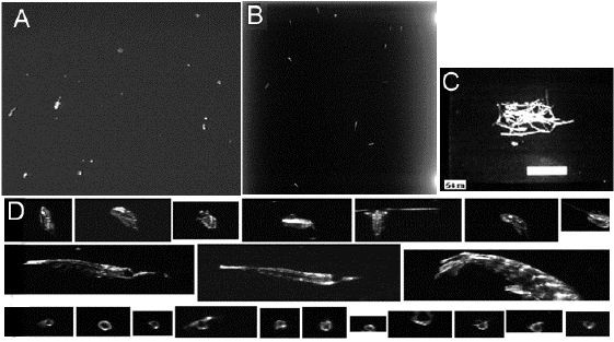
A: Particle image from with a structured lighting, high
resolution color video camera system (Pilskaln et al, 1998). B: Image
collected with the zooplankton visualization and imaging system (ZOOVIS)
designed to collect high resolution images of meso- and macrozooplankton
from relatively large volumes of water (102-103 mls) (Benfield et al.
2002). Image contains at least 13 euphausiids. C: Algal mat image from No.
Pacific obtained with a towed VPR with an analogue black and white video
camera and synchronized strobe (Darkangelo et al. 1996; Villareal et al.
1999). White bar is 1 cm. D: Zooplankton images collected with a VPR.
Digitized video fields are scanned by the software for targets (referred
to as regions of interest, ROI s) which meet previously defined criteria
of brightness, focus and size. ROIs are classified visually into taxonomic
groups by examination of each image in a thumbnail browsing program (Benfield
et al. 1996). Top row: Calanus finmarchicus copepods; middle row: complete
and partial images of euphausiids likely Meganyctiphanes norvegica; and
bottom row: Limacina retroversa pteropods. These represent 3 of maybe 20
or 30 possible classes encountered in a study.